
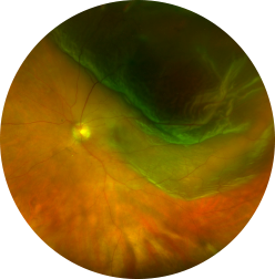
What is a Retinal Detachment?
The retina is the light-sensitive layer of nerve tissue that lines the inside of the eye and sends visual messages through the optic nerve to the brain. A retinal detachment occurs when the retina becomes separated from the rest of the layers of the eye. This usually occurs after you develop a tear in the retina. The extent of permanent damage depends on how much of the retina becomes detached and whether or not the center of the retina (the macula) becomes detached. The macula is made up of special nerve cells that provide the sharp central vision needed for seeing fine detail (reading, driving etc.). If your macula has become detached, you have a poor visual prognosisand you may not regain good enough vision to read or drive with that eye even after successful surgery.
Why do I have a Retinal Detachment? What are the symptoms?
A retinal detachment occurs when a tear forms in the retina allowing fluid to get under the retina forming a detachment. They are more common in patients who are very near-sighted, have a family history of retinal detachment, and in eyes that have had prior trauma or eye surgery. Patients often complain of flashes, new floaters and a shadow forming in their vision when a retinal detachment occurs.
Assessment for Retinal Detachment
We are able to detect a retinal detachment during an eye examination. Your surgeon will carefully examine your eye to identify all the retinal tears and determine the extent of the retinal detachment. He may need to press on your eye to examine your retina fully. He will then discuss with you an appropriate surgical plan to most safely and effectively reattach your retina.
There are three basic options to repair of a retinal detachment: