
What is a Macular Hole?
A macular hole is a condition where a very small hole has developed right in the center of the retina in an area that is responsible for our sharpest vision. The part of the eye affected is called the macula. The macula is made up of special nerve cells that provide us with the sharp central vision we need for seeing fine detail (reading, driving etc.). When a macular hole develops, you will suffer from symptoms such as a dark area right in the center of your vision, distortion, or general blurring.
Why do I have a Macular Hole?
We do not know why people develop macular holes. However, we do know that it occurs more often in patients later in life (60’s and older) and occurs more often in females. Macular holes are not related to macular degeneration. If you have a macular hole you have less than a 25% risk of developing a macular hole in your fellow eye.
Assessment for Macular Hole:
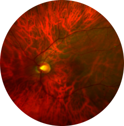
We are able to detect a macular hole during an eye examination. Sometimes, a special scan of the back of the eye (Optical Coherence Tomography) may be needed to confirm the presence of a macular hole. If a macular hole is present, your surgeon will likely recommend a surgical procedure to try to close the hole and improve your vision. Surgery is the only way to treat a macular hole; there are no eye drops or medications that you can take that will help.
Outcomes of Macular Hole Surgery:
Although a majority of our patients experience improved vision after surgery, there are a small percentage of patients that do not have an improvement in vision even after successful and uncomplicated surgery. Patients that do not have a significant improvement in vision after surgery despite closure of the macular hole often have had their macular hole for a longer period of time (greater than 1 year). If you elect to proceed with macular hole surgery, there is a 95% chance of macular hole closure with one surgery. This assumes that you comply with face down positioning after the surgery. You need to keep in mind that our goal is to maximize the vision in your affected eye. Even after successful surgery, your vision will likely never be as good as it was prior to the onset of your symptoms.
Risks of Macular Hole Surgery:
There are several risks associated with macular hole surgery that you need to be fully aware of prior to electing to proceed with surgery. The most common are as follows:
The Surgical Procedure:
Macular hole surgery involves a vitrectomy and membrane peeling. We use the most advanced surgical equipment and techniques available for macular hole surgery. The surgery involves making small holes in the eye and using instruments to remove the jelly-like substance that normally fills the back chamber of the eye, called the vitreous (vitrectomy). The vitreous is replaced naturally by fluid produced inside the eye. A thin membrane on the surface of the retina surrounding the macular hole is then physically peeled off the retina with a fine forceps (membrane peeling). The eye is then filled with an inert gas which will slowly diffuse out of the eye over 4-6 weeks. The holes made in your eye are made in a fashion that allows them to close and heal on their own; usually no stitches are needed to close them. The surgery usually takes less than one hour to perform. It is typically performed under general anesthesia.
What should I expect following surgery?
After the surgery you will have an eye dressing placed on your eye. Once you get to the recovery room you will be placed in a face-down position. Do not remove your eye shield until we see you in the clinic the following day when we will remove it for you. You will be given instructions on which eye drops to use and all your restrictions at this time. You do not need to use any eye drops the night after surgery. Please bring all your eye drops to this first postoperative visit.Most patients have minimal discomfort after surgery. When your eye shield is taken off your vision will typically be more blurred because of the gas bubble in your eye. Your vision will improve once the gas bubble dissipates in 4-6 weeks.
Face Down Positioning:
It is very important that you position face down for the amount of time advised by your surgeon. You need to be in this position so the gas bubble remains in the appropriate position to help the macular hole close. If you do not position your macular hole may not close. We can provide you with information on renting equipment that patients often use to make face down positioning more comfortable and easier.